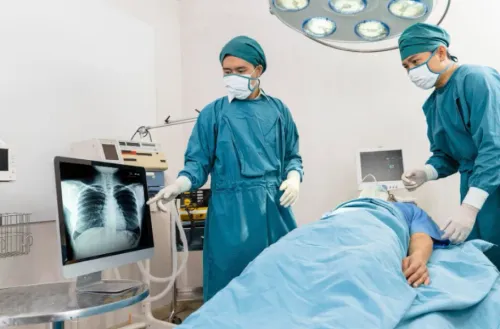
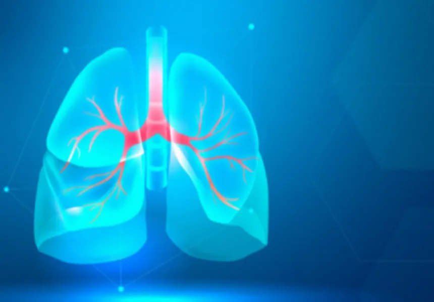
The term lung defects can describe several congenital, structural, or acquired abnormalities affecting the lungs and surrounding structures. Some are identified early in life, while others may remain unnoticed until adulthood or appear following infection, injury, or another disease.
Understanding the nature of an abnormality is important before deciding whether monitoring, medical treatment, or surgery is appropriate.
Different Types of Structural Lung Problems
Not every lung abnormality has the same cause or clinical significance.
A patient may be evaluated for:
- Congenital abnormalities
- Localised structural changes
- Damaged lung tissue
- Abnormal air-filled spaces
- Pleural abnormalities
- Persistent cavities
- Airway-related structural problems
- Abnormal findings discovered on CT imaging
Some findings are harmless or require only observation. Others can contribute to recurrent infection, breathing difficulties, bleeding, or progressive loss of lung function.
Symptoms Depend on the Condition
Structural abnormalities do not produce one universal symptom pattern.
Possible concerns include persistent cough, repeated chest infections, shortness of breath, chest discomfort, coughing up blood, unexplained reduction in exercise tolerance, or recurrent respiratory symptoms.
However, many of these complaints can occur with common respiratory illnesses. Imaging and clinical assessment are therefore essential for identifying the underlying cause.
Why CT Imaging Is Often Important
A chest X-ray can provide useful initial information, but CT imaging offers a more detailed view of the lungs and surrounding anatomy.
A CT scan can help doctors assess:
- Location of an abnormality
- Size and extent
- Relationship to nearby structures
- Changes in lung tissue
- Presence of fluid or air
- Features requiring further investigation
In selected cases, additional tests such as bronchoscopy, pulmonary function testing, biopsy, or specialised imaging may be needed.
When Is Surgery Considered?
Surgery is generally considered when there is a clear clinical reason for intervention.
This may include an abnormality causing repeated complications, suspicious tissue requiring removal, localised irreversible damage, persistent air leakage, or another structural problem that can be corrected surgically.
The goal is not simply to remove abnormal tissue. Surgeons must balance treatment of the condition with preservation of healthy lung function.
Lung Function Matters Before an Operation
Before major lung surgery, doctors often evaluate how well the lungs currently work.
Pulmonary function tests can help estimate respiratory reserve. This information becomes particularly important if a significant amount of lung tissue may need to be removed.
Smoking history, asthma, chronic obstructive pulmonary disease, previous infections, age, physical capacity, and cardiac health may also influence surgical planning.
Why Cardiothoracic Assessment Can Be Useful
The lungs operate inside the chest alongside the heart, major blood vessels, airways, and other structures. Complex abnormalities may therefore require expertise extending beyond routine respiratory treatment.
A thoracic or cardiothoracic surgeon can assess whether an anatomical problem has a surgical solution and explain the potential benefits and limitations of intervention.
Patients in Pune who have received an unexpected CT finding should bring previous scans and reports to their consultation whenever possible. Comparing current imaging with older studies can reveal whether an abnormality has remained stable or changed over time.
Dr Ashish Khanijo Cardiac Specialist provides assessment for selected cardiothoracic conditions involving the heart, lungs, chest, and major blood vessels.
A diagnosis described simply as a lung defect should always be clarified. Patients benefit from knowing the exact medical name of the condition, its likely cause, whether it is progressing, how it affects lung function, and what treatment options are realistically available.
Keywords